
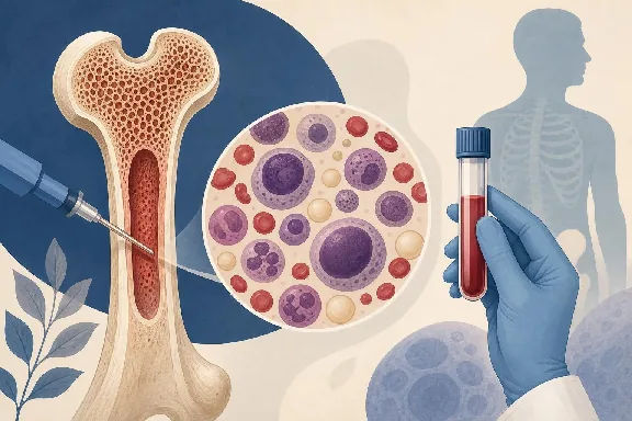
Learn what a bone marrow test shows and the conditions it can detect, from blood cancers to infections, to gain clarity on your health and diagnosis.
When your doctor mentions a bone marrow test, it’s natural to feel a wave of anxiety. But understanding what this procedure reveals can replace fear with clarity. A bone marrow test shows how well your body produces blood cells: red cells that carry oxygen, white cells that fight infection, and platelets that stop bleeding. It can detect conditions ranging from blood cancers to hidden infections, and it often provides answers that standard blood tests simply can’t. Whether your doctor suspects a serious disease or wants to rule one out, this test offers a direct look at the factory where your blood is made.
The results can shape treatment decisions, confirm or exclude diagnoses, and sometimes reveal problems no one expected. If you or someone you care about is facing this procedure, knowing what it can uncover helps you ask better questions and feel more prepared for what comes next. Here’s a straightforward breakdown of the conditions a bone marrow test can identify and what the results actually mean.
Understanding Bone Marrow Biopsy and Aspiration
Bone marrow testing isn’t a single procedure: it’s typically two complementary techniques performed during the same visit. Both target the spongy tissue inside your bones, usually the back of the hip bone (posterior iliac crest), because that site gives the most representative sample. The entire process generally takes 15 to 30 minutes, and most patients go home the same day.
The Difference Between Aspiration and Biopsy
An aspiration uses a hollow needle to withdraw a small amount of liquid marrow. This fluid sample lets pathologists examine individual cells under a microscope, checking their size, shape, and maturity. They can also run flow cytometry and genetic tests on this liquid sample.
A biopsy, on the other hand, removes a tiny core of solid bone and marrow tissue, usually about 1 to 2 centimeters long. This core preserves the architecture of the marrow: how cells are arranged, how dense they are, and whether anything abnormal is growing in the tissue structure. Doctors almost always perform both together because each provides information the other can’t.
Why Doctors Order Bone Marrow Testing
Your hematologist won’t order this test on a whim. It’s typically requested when blood work shows persistent abnormalities: unexplained anemia, abnormal white cell counts, unusual cells circulating in the bloodstream, or platelets that are too high or too low without a clear cause. Sometimes it’s ordered to stage a cancer already diagnosed through other means, or to check whether a treatment like chemotherapy is working.
Bone marrow testing is also used before stem cell transplants to evaluate donor compatibility and marrow health. In some cases, it’s the only way to confirm a diagnosis that blood tests can suggest but never prove on their own.
Detecting Blood Cancers and Malignancies
One of the primary reasons doctors order bone marrow tests is to look for cancer. Blood cancers often originate in or spread to the marrow, making this test essential for accurate diagnosis and staging.
Leukemia and Lymphoma Diagnosis
Leukemia starts in the bone marrow itself, so examining marrow tissue is the gold standard for diagnosis. The test reveals the type and percentage of blast cells (immature, abnormal cells) present. In a healthy adult, blasts make up less than 5% of marrow cells. When that number climbs above 20%, it typically confirms acute leukemia.
Different types of leukemia: acute myeloid (AML), acute lymphoblastic (ALL), chronic myeloid (CML), and chronic lymphocytic (CLL) each produce distinct patterns that pathologists can identify. For lymphoma, marrow biopsy determines whether the cancer has spread beyond the lymph nodes. This staging information directly affects treatment planning and prognosis.
Multiple Myeloma and Plasma Cell Disorders
Multiple myeloma is a cancer of plasma cells, which normally produce antibodies. A bone marrow biopsy can quantify the percentage of abnormal plasma cells in the marrow. Healthy marrow contains roughly 2-3% plasma cells; myeloma patients often show 10% or more, sometimes exceeding 60%.
The test also helps identify precursor conditions like monoclonal gammopathy of undetermined significance (MGUS) and smoldering myeloma. Distinguishing between these stages matters enormously because MGUS may only need monitoring, while active myeloma requires treatment.
Monitoring Cancer Progression and Treatment Response
Bone marrow tests aren’t just for initial diagnosis. Oncologists use repeat biopsies to track how well treatment is working. After chemotherapy, the test can show whether cancer cells have been eliminated, reduced, or are persisting despite treatment.
The concept of measurable residual disease (MRD) has become increasingly important in 2026\. Using highly sensitive techniques like flow cytometry and next-generation sequencing on marrow samples, doctors can detect as few as one cancer cell among a million normal cells. This level of precision helps guide decisions about continuing, changing, or stopping therapy.
Identifying Non-Cancerous Blood Disorders
Cancer gets the most attention, but bone marrow tests detect a wide range of non-cancerous conditions too. These disorders can be just as debilitating and often require their own specific treatments.
Anemia Types and Iron Stores
Not all anemia is the same, and a bone marrow test can differentiate between types that look similar on standard blood work. Iron-deficiency anemia, sideroblastic anemia, and anemia of chronic disease each leave distinct fingerprints in the marrow.
One particularly useful finding is the Prussian blue stain, which reveals iron stores within marrow cells. If iron stores are absent, it confirms iron deficiency. If iron is present but trapped inside abnormal ring-shaped formations around the cell nucleus (ring sideroblasts), it points toward myelodysplastic syndrome or sideroblastic anemia. This distinction changes treatment entirely: iron supplements won’t help if the problem is how cells process iron rather than how much is available.
Aplastic Anemia and Bone Marrow Failure
Aplastic anemia occurs when the marrow stops producing enough blood cells. A biopsy is the definitive way to diagnose it because it shows a marrow that’s been replaced by fat cells instead of the blood-forming tissue that should be there. Normal marrow cellularity in a young adult is around 60-70%; in severe aplastic anemia, it can drop below 25%.
This diagnosis carries significant weight because treatment options range from immunosuppressive therapy to bone marrow transplant. The biopsy also helps rule out other causes of pancytopenia (low counts of all blood cell types), such as myelodysplastic syndromes or hairy cell leukemia, which can mimic aplastic anemia on blood tests alone.
Polycythemia Vera and Thrombocytopenia
Polycythemia vera is the opposite problem: the marrow produces too many red blood cells, and often too many white cells and platelets as well. A bone marrow biopsy shows hypercellular marrow with increased megakaryocytes (platelet-producing cells) and characteristic clustering patterns. Combined with JAK2 mutation testing, the biopsy helps confirm the diagnosis according to 2022 WHO criteria still used in clinical practice.
For patients with unexplained low platelet counts, a marrow test can distinguish between conditions where platelets are being destroyed in the bloodstream (like immune thrombocytopenia) versus conditions where they aren’t being produced adequately. This distinction is critical because the treatments are fundamentally different.
Diagnosing Infections and Storage Diseases
Beyond blood cell disorders, bone marrow tests can uncover infections and rare metabolic conditions that are difficult to diagnose through other means.
Hidden Infections in the Bone Marrow
Certain infections hide within the marrow, making them nearly invisible to standard diagnostic methods. Tuberculosis, histoplasmosis, and leishmaniasis can all be identified through marrow examination when cultures and imaging come up empty. In immunocompromised patients, including those with HIV or those on immunosuppressive medications, disseminated fungal infections sometimes show up first in marrow samples.
Granulomas, which are clusters of immune cells responding to infection, can appear on biopsy and prompt further investigation. The marrow sample can also be cultured for bacteria, fungi, and mycobacteria, providing another avenue for identifying the specific organism causing illness.
Metabolic and Genetic Storage Disorders
Rare conditions like Gaucher disease, Niemann-Pick disease, and other lysosomal storage disorders leave distinctive cells in the marrow. Gaucher disease, for instance, produces characteristic "Gaucher cells": large macrophages with a crinkled-tissue-paper appearance that pathologists can spot under the microscope.
These storage disorders involve enzyme deficiencies that cause certain substances to accumulate in cells. While genetic testing has become the primary diagnostic tool for many of these conditions, marrow examination still plays a role in initial discovery, especially when the condition wasn’t suspected and the characteristic cells are found incidentally during a biopsy performed for other reasons.
Interpreting Your Bone Marrow Results
Getting the results is one thing; understanding them is another. Bone marrow reports are dense and technical, but knowing the basics helps you have a more productive conversation with your doctor.
Cellular Morphology and Genetic Testing
The pathology report typically includes several components. The morphology section describes what cells look like: their size, shape, maturity, and any abnormalities. You’ll see terms like "normocellular" (normal cell density), "hypercellular" (too many cells), or "hypocellular" (too few).
Beyond what’s visible under a microscope, modern bone marrow analysis includes:
- Flow cytometry: identifies cell types by their surface markers, crucial for classifying leukemias and lymphomas
- Cytogenetics: examines chromosomes for deletions, translocations, or extra copies that indicate specific diseases
- FISH (fluorescence in situ hybridization): detects targeted genetic abnormalities faster than standard cytogenetics
- Molecular testing: identifies specific gene mutations like JAK2, BCR-ABL, or FLT3 that guide treatment choices
These tests run simultaneously on your sample, each answering different diagnostic questions.
Timeline for Receiving Results
Don’t expect everything at once. Preliminary morphology results from the aspiration may be available within 1 to 3 days. The biopsy tissue, which needs to be processed and sectioned, typically takes 3 to 7 days.
Genetic and molecular results take longer: cytogenetics usually require 10 to 14 days because cells need to grow in culture before chromosomes can be analyzed. Molecular tests like next-generation sequencing panels may take 2 to 3 weeks. Your hematologist will often share preliminary findings while waiting for the complete picture, especially if urgent treatment decisions need to be made.
What This Means for You
A bone marrow test is one of the most informative diagnostic tools in medicine. It reveals conditions that blood tests can only hint at, from leukemia and myeloma to aplastic anemia, hidden infections, and rare genetic disorders. The information it provides often determines not just a diagnosis but the specific subtype and genetic profile of a disease, which directly shapes treatment.
If your doctor has recommended this test, ask about what they’re specifically looking for and what the possible outcomes mean for your care plan. Understanding what a bone marrow test can show gives you the foundation to participate meaningfully in those conversations. The procedure itself is brief, and while it’s not comfortable, the diagnostic clarity it provides is often irreplaceable. Whatever the results reveal, they bring you closer to answers and, ultimately, to the right treatment.